The testing may vary slightly for each patient depending on age, condition, patient’s preference.
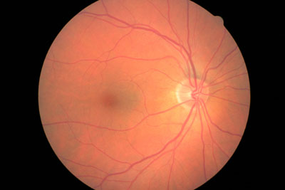
Our NIDEK Digital Retinal Camera takes a digital image of the back of the eye, and so gives us the opportunity to detect, view and monitor early signs of any
sight-threatening eye conditions.
If required, the digital images of the retina can then be emailed to a doctor or ophthalmologist after your eye exam. Our
state of the art retinal camera helps the early detection of problems such as macular degeneration, glaucoma, retinal detachments and diabetic
retinopathy. These conditions commonly affect hundreds of thousands of people in Canada; the sooner they are detected, the sooner we can suggest a
solution.
The traditional ophthalmoscope does not take a picture of the eye, so after the eye examination, there is no visual record of its condition. In contrast, digital retinal
photography gives us a permanent image of the eye which can then be compared to images taken in future years.
Detect sooner. Protect more.
With certain eye conditions, the faster you act, the more you protect. That’s why we recommend OCT as part of your regular eye
exam. It can help us detect sight threatening issues up to 5-10 years before traditional testing methods, and many years before you notice any change in your
vision yourself.
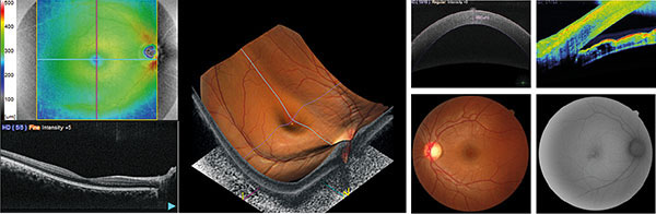
What is OCT?
Optical Coherence Tomography (OCT) is a non-invasive test that uses light waves [no radiation] to show cross-sectional pictures of the different retinal layers and works
a little like ultrasound for the eye – allowing us to spot abnormalities up to 5-10 years before a traditional test.
(Click here to learn more about other common eye conditions.)
The images generated by the OCT are stored on your file so when you see us again for your next appointment, the images from the latest screening can be compared with
previous images to see if anything has changed or deteriorated.
The sooner we detect, the sooner we can help you treat.
Why have an OCT screening?
Spot issues sooner – an OCT scan allows us to detect and monitor any changes to your eye health up to 5-10 years earlier than other methods of eye
screening.
See more – we can instantly show you the 3D High Definition images of your eyes.
Build a more complete picture – all your scans are stored on your records. So from test to test, we can compare your OCT images, and quickly pinpoint
any changes in your eye health.
The OCT examination is completely non-invasive and painless. Nothing touches your eye at all. It only takes a few seconds, after that the images generated are examined
by your optometrist.
What can you see?
The OCT screening generates a series of 3D High Definition (HD) scans – just like the one below (click on images in enlarge):
We recommend you have them as part of your regular eye examination. That way you have ultimate peace of mind about your eye health.
How do I book it?
We are accepting new patients, no referrals necessary.
The eye uses central vision for “sight” to identify clarity and detail; and peripheral vision to assess our orientation in the world around us and to detect movement.
Unfortunately, peripheral vision can be reduced or lost in conditions such as glaucoma and other optic nerve diseases, but also from damage to visual pathways, e.g.
certain brain tumours, stroke. This loss is irreversible and may not be noticed until it is quite severe.
A.k.a. “the clicky test” it repeatedly presents a light spot in different areas of your peripheral vision.
The Zeiss VFA is known as Gold standard of perimetry worldwide. It tests the range and quality of your peripheral vision to identify any loss sooner and protect the
vision. It is also used in combination with other testing to diagnose the severity and progression of disease.
Most people have had a traditional phoropter placed in front of your eyes to help the Optometrist select the correct prescription through a series of tests and lens
changes. This automated version however allows for a smoother swifter test; and once your prescription is determined, our Doctor can instantly show you the comparison
with your unaided eye, your current eyeglasses and the new prescription.
Before the Optometrist determines your ideal prescription, we use a computerised pre-screening instrument to obtain a starting point. This automatic result is then fine
tuned into an exact prescription that will work best for you. Our prescreening equipment allows us to perform 4 tests-in-1, saving time and reducing the inconvenience
of moving from one instrument to another. So, the corneal curvature and thickness and intraocular pressure are also measured from the same machine.
At Royal London Optometry, we utilise the latest technology from the UK in the form of a Computerised Test Chart.
The flat panel display has the most comprehensive range of vision assessment tools from letters, numbers, pictures, different languages, images for fixation including
photographs and cartoons including our very own Coco the Clinic Clown!! In addition, it has great patient education animations and videos that help
explain conditions in a more patient friendly manner.
Legal Notice
The information below applies to all the information contained on the royallondonoptometry.ca Website.
The users of this Website agree to comply with the terms set out below.
TERMS OF USE
Royal London Optometry owns and operates a Website at royallondonoptometry.ca (hereinafter referred to as the “site” or
“Website”). Unless specified otherwise on the site, it is accessible to all users (hereinafter, the "user"). Refrain from using this Website unless you
agree to comply with the conditions.
RISKS ASSOCIATED WITH INFORMATION
Information on this Website is intended for informational purposes only and has no contractual value.
Royal London Optometry reserves the right to modify the content of this site. At any time, without prior notice.
Royal London Optometry assumes no liability for errors or omissions in the content of this Website or for
information reliability or completeness of said information.
Information published on this Website is based on marketing, statistical or commercial services or other sources
the Royal London Optometry considers reliable and are the sole responsibility of their authors and not of
Royal London Optometry. We do not assume any liability for the accuracy or completeness of said information and in
no circumstance should this information be regarded as such. Opinions and information as presented on this site
reflect our position as of the date of publication and are subject to change without notice.
UPDATING OF THE WEBSITE UNDER THE RESPONSIBILITY OF Royal London Optometry
Royal London Optometry, its employees and directors will not be liable for damages incurred as a result of the
information published on this site, for the views and advice published, expressed or implied regardless of its
nature.
Royal London Optometry expressly refuses any and all responsibility for the manner in which the user of the site may
use the information contained, in any decisions that may be made and in the actions that may or may not be taken
based on said information.
COPYRIGHT PROTECTION
Presentations made and contained on this site are the intellectual property of Royal London Optometry. Reproduction
in whole or in part of this site on any other medium in prohibited without the express permission of
Royal London Optometry.
User may solely use the information contained on this site for personal use. Reproduction in whole or in part of
said information on paper may only be performed for personal use. Said information is not to be copied,
distributed or transmitted to third parties nor may it be inserted in a document or other medium.
HYPERTEXT LINKS
The links to external Websites and their content shall not be prejudged and Royal London Optometry will in no way be
held responsible for any direct or indirect prejudice that may result from gaining access to and usage of said
sites.
CONFIDENTIALITY
Royal London Optometry draws the attention of the user to the fact that all communication transmitted through this
Website remains in the public domain and not the private domain. Royal London Optometry cannot accept responsibility for the
security of the transmission of information.
The confidentiality and integrity of the information circulating over the internet cannot be ensured.
Royal London Optometry cannot accept responsibility in the case that data contained on this site is intercepted.
Royal London Optometry site uses cookies. These cookies are small text files saved on the hard disk of a user's
computer. These files are completely harmless and cannot contain viruses. These cookies are used to analyze
visits to the site. Royal London Optometry calls upon Google Analytics to help track how users use the site. The
number of visitors, path taken to access the site and length of each visit are measured. The cookies cannot, in
any way, identify the user. All data is completely anonymous and compiled solely for the purpose of improving
the site and tailoring the content to the needs of its visitors.
The person responsible for the protection of personal information is the owner of : Royal London Optometry
VIRUSES AND TECHNICAL GLITCHES
Royal London Optometry makes no representations that the content of this site is free of infections, viruses, worms,
Trojan horses and/or other codes with contaminating or destructive properties. It is the user's responsibility
to take protective measures.
Royal London Optometry DECLINES ALL RESPONSIBILITY IN THE EVENT OF ANY INTERRUPTION OR NON-AVAILABILITY OF THE
SERVICE
Under no circumstances shall Royal London Optometry be held responsible for transmission errors of any sort, such as
loss of or damage to data, or changes of any type whatsoever, including direct or indirect damage resulting from
the use of the services provided on this site.
JURISDICTION
This Agreement shall be governed by and construed in accordance with the laws of the province of BC.
Any dispute arising of this Agreement shall be brought before the court in the judicial district of
Royal London Optometry's head office. Address: 2 - 3248 King George Blvd, Surrey BC; Tel: [778] 294-2236.
PUBLISHING FIRM
Royal London Optometry
2 - 3248 King George Blvd, Surrey BC Tel: [778] 294-2236
Acceptance of the Privacy Policy
Thank you for visiting royallondonoptometry.ca (the “Website”), provided to you by Royal London Optometry (“We”). We
respect the privacy of every individual who visits the Website and are sensitive to privacy
issues on the Internet. We believe it is important that you know how we deal with information
received about you.
This privacy policy (the “Privacy Policy”) explains how we collect, use, disclose, and protect
the personal information of our customers and Website users ("you"), describes the types of
information we may collect from you or that you may provide to us, and our practices for
collecting, using, maintaining, protecting, and disclosing that information. The Website is for
general audiences and is not specifically targeted to or intended for use by children.
We will only use your personal information in accordance with this Privacy Policy unless
otherwise required by applicable law. We take steps to ensure that the personal information that
we collect about you is adequate, relevant, not excessive, and used for limited purposes.
Privacy laws in Canada generally define "Personal Information" as any information about an
identifiable individual, which includes information that can be used on its own or with other
information to identify, contact, or locate a single person.
By accessing or using the Website, you are accepting the practices described in this Privacy
Policy, and you are consenting to our processing of your information as set out in this Privacy
Policy. We may modify or update this Privacy Policy from time to time; if we change this Privacy
Policy in a manner that materially impacts your privacy rights, we will provide a notice to you.
Your continued use of the Website or our services after any modification to this Privacy Policy
will constitute your acceptance of such modification. However, when required by law, we will
confirm your consent to the revised Privacy Policy terms. This Privacy Policy is incorporated
into and considered a part of the Website Terms and Conditions of Use, located here
What information we collect
We collect and use several types of information from and about you, including:
Personal Information, that we can reasonably use to directly or indirectly
identify you, such as your full name, email address, telephone number and any other
identifier we may use to contact you online or offline.
Non-personal information is information that does not directly or
indirectly reveal your identity or directly relate to an identified individual, such as
demographic information, or statistical or aggregated information. Statistical or aggregated
data does not directly identify a specific person, but we may derive non-personal
statistical or aggregated data from Personal Information. For example, we may aggregate
Personal Information to calculate the percentage of users accessing a specific Website
feature.
How we collect the information
Information You Provide to Us
The information we collect directly from you may include:
Communicating with us: When you contact us about our services or to make other inquiries, we collect the content of those
communications, as well as your full name, email, phone number, and any additional information that allows us to answer your request.
Information We Collect While You Interact With Us Through Cookies and Other Automatic Data
Collection Technologies
We use your information, including your Personal Information, to manage our business and to
maintain and develop commercial relationships with you. We will collect, use, and disclose such
information only to the extent that is necessary for those purposes.
We use information that we collect about you or that you provide to us, including any Personal
Information:
To provide you with information, products, or services that you request from us.
To fulfill the purposes for which you provided the information or that were described when
it was collected, or any other purpose for which you provide it.
To improve the Website, products or services, marketing, or customer relationships and
experiences.
To allow you to participate in interactive features, social media, or similar features on
the Website.
To measure or understand the effectiveness of the advertising we serve to you and others,
and to deliver relevant advertising to you.
In any other way we may describe when you provide the information.
For any other purpose with your consent.
Partnership with Microsoft Clarity and Microsoft Advertising
We may partner with Microsoft Clarity and Microsoft Advertising to capture how you use and interact with our website through behavioral metrics, heatmaps, and session replay to improve and market our products/services. Website usage data is captured using first and third-party cookies and other tracking technologies to determine the popularity of products/services and online activity. Additionally, we use this information for site optimization, fraud/security purposes, and advertising. For more information about how Microsoft collects and uses your data, visit the Microsoft Privacy Statement.
How we share your information
We will not rent or sell your information to third parties without your consent. We only share
your data as specifically provided in this Privacy Policy.
Other Disclosures
In addition to any disclosure you may have consented to or permitted under the terms of this
Privacy Policy, we may transfer your data, including Personal Information to third parties in
the following limited circumstances:
information you expressly consent to be shared;
when relating to anonymized information (individuals cannot be identified by it);
when you decide to make the information indexable by search engines, to share or to
distribute the information to people or otherwise to make it available to the public;
to satisfy any applicable law, regulation, legal process or enforceable governmental request
within or outside your country of residence when we have a good faith belief that the law
requires it;
to enforce this Privacy Policy, the Terms, or an agreement, including investigation of
potential violations thereof;
to detect, prevent, or otherwise address fraud, security or technical issues, or protect the
operations or you;
to protect our rights, property or safety as well as yours, the public, or others;
in connection with an acquisition, merger, change in control, debt financing,
reorganization, sales of assets, bankruptcy or other change of our corporate structure or
status; or
as necessary in connection with the performance of requested services or solutions, or as
otherwise appropriate in connection with a legitimate need.
How we store and secure your information
Information Security
We are committed to protecting the confidentiality, integrity, availability and privacy of your Personal Information. We have put appropriate physical, technological and procedural security measures in place designed to help prevent your Personal Information from being lost, used, modified or accessed in an unauthorized way, or improperly disclosed. Examples of such measures include restricted access to offices, training of personnel, using passwords and well-defined internal policies and practices. We also use encryption technology and Secure Socket Layers ("SSL") in all areas of the Website where your personal account information is required.
In addition, we limit access to your Personal Information to those employees, agents, contractors and other third parties who have a business need-to-know. They will be required to process your Personal Information only on our instructions and they are subject to an obligation of confidentiality. Our service providers are required to maintain adequate security protections in place designed to help safeguard your Personal Information and are not permitted to use it for any purpose other than fulfilling services to us.
If you have any questions about securing your personal data, please contact us in accordance with the “How to Contact Us” section below.
Information Retention
We will retain your Personal Information for as long as it is needed:
to provide the products and services that you have requested;
to communicate with you about a purchase or a request you have made to us;
to manage your choices and rights you have exercised pursuant to this Privacy Policy;
to comply with our legal and regulatory obligations and to demonstrate compliance,
to resolve disputes and to enforce our rights and agreements.
We may retain non-personal information that has been sufficiently aggregated or anonymized for a longer period.
Once the retention period is over, we will dispose of your Personal Information as provided for in our internal data retention and disposal policy.
Where We Store Information
We use facilities operated by "Amazon Web Services" and located in Canada as our information storage and processing infrastructure. Our service providers can also, from time to time, store your Personal Information in accordance with purposes outlined in this Privacy Policy.
How to contact us
If you have any questions about this Privacy Policy, you can contact us at [778] 294-2236.
Cookie Policy – royallondonoptometry.ca
This Cookie Policy explains what cookies are and how we use them. You should read this Policy to
understand what type of cookies we use, the information we collect from the cookies, and how
that
information is managed. For further information on how we handle, store and keep your personal
data
secure, see our Term of use.
What are Cookies?
Cookies are small text files which are stored on the browser or hard drive of your computer or
mobile
device when you visit a webpage or application. A “session cookie” stores temporary information
that
is deleted when you close your web browser or turn off your computer or device. A “persistent
cookie” enables the site to recognize when you return to it and remains stored on your computer
until you delete it. Cookies work to make your experience browsing our site as smooth as
possible by
remembering your preferences the next time you visit the site.
What types of cookies do we use?
There are four variety of cookies which operate on our website:
Essential cookies allow you to be able to experience the full functionality
of
our site. Without these cookies, some parts of our site may not work as they
should.
Performance cookies tell us how you use our site and help us to improve it.
For
example, these cookies count the number of visitors to our website and see how visitors move
around when they are using it. This helps us to improve the way our site works, for example,
by
ensuring that users find what they are looking for easily. The information collected by
these
cookies is anonymous.
Google Analytics is a performance cookie used to track visits to
our
website and user behaviour on site. The data collected is anonymous and is used for
benchmarking purposes to monitor the performance of our site and to measure user
engagement on our site.
Social media cookies from social media sites such as
Facebook, Instagram, and
Pinterest
are used to enable social media buttons on our site to work. Social media buttons
allow
you to share content and interact with your social network. These social media
platform
may set their own cookies on your device. We do not control the settings of these
cookies so we suggest you check the social media website for more information about
their cookies and how to manage them. Our website will not collect or store any
personally identifiable information from the user. To find out how you can opt out
of
accepting these cookies, please visit the cookie policy and privacy policies of the
social media site.
Customizer cookies allow our site to remember your preferences, helping to
customize your experience on our site.
Our customizer cookie, which helps us to manage the technology and cookies which are
used across our site.
Targeting/advertising cookies are set by third party advertising partners
through our digital touchpoints to build profiles based on your interests. These cookies
enable
us to send you relevant content and advertising based on your preferences and track the
effectiveness of our ad campaigns.
Below are several advertising and targeting cookies that are used to identify
different
browsers and devices. They collect data anonymously for the purposes of ad
selection,
reporting, behavioural targeting, and cross-device advertising (i.e. associating
multiple devices together). In some instances, we may provide a hashed version of
your
email address or other information to the platform provider for such proposes. More
information can be found at the following links.
How do you change cookie preferences or block cookies?
Within your browser, you can choose whether you wish to accept cookies or not. Different browsers
make different controls available to you. Generally, your browser will offer you the choice to
accept, refuse or delete cookies at all times, or those from providers that website owners use
i.e.
third party cookies, or those from specific websites. Each browser’s website should contain
instructions on how you can do this.
If you block cookies on our website, you may be unable to access certain areas of our website and
certain functions and pages will not work in the usual way.
Changes to the Cookie Policy
We may update this Policy from time to time. If we make significant changes we will let you know
but
please regularly check this Policy to ensure you are aware of the most updated version.
This Cookie Policy was last updated on February 10, 2025.
We have successfully received your message and will get back to you as soon as possible.
An unexpected error occurred. Please try again later.